Antigens
| Title | Description | Image |
|---|---|---|
| CD057 (Leu 7) | (membraneus en cytoplasmatisch) Tonsil: positieve NK cellen en geactiveerde Tcellen (subsets) in het kiemcentrum en in de T cel zone zijn sterk aangekleurd | 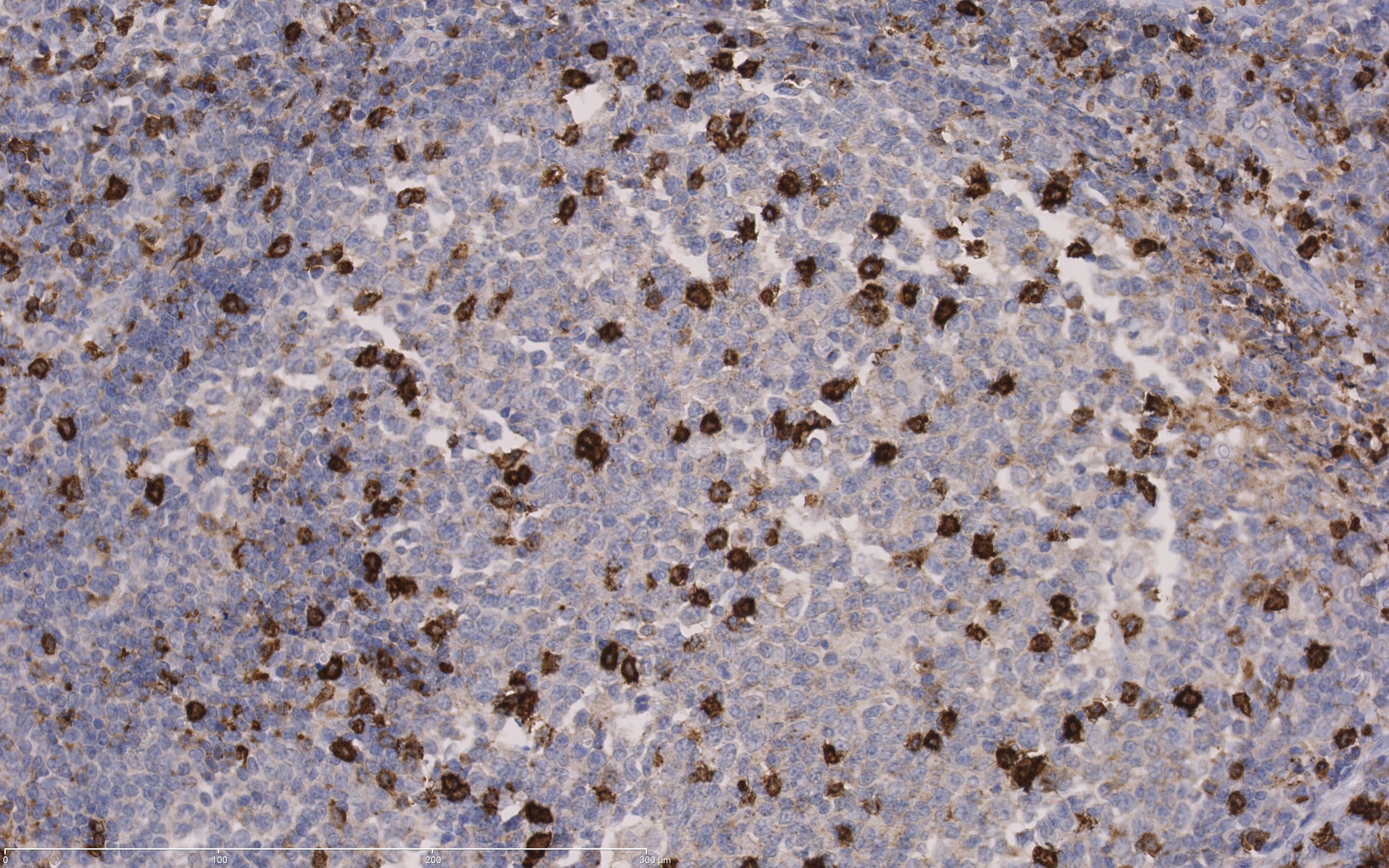
Tonsil: CD57 positive NK cells and T-cell subsets
|
| CD061 (GP IIIa) | Beenmerg: megakaryocyten zijn positief. | 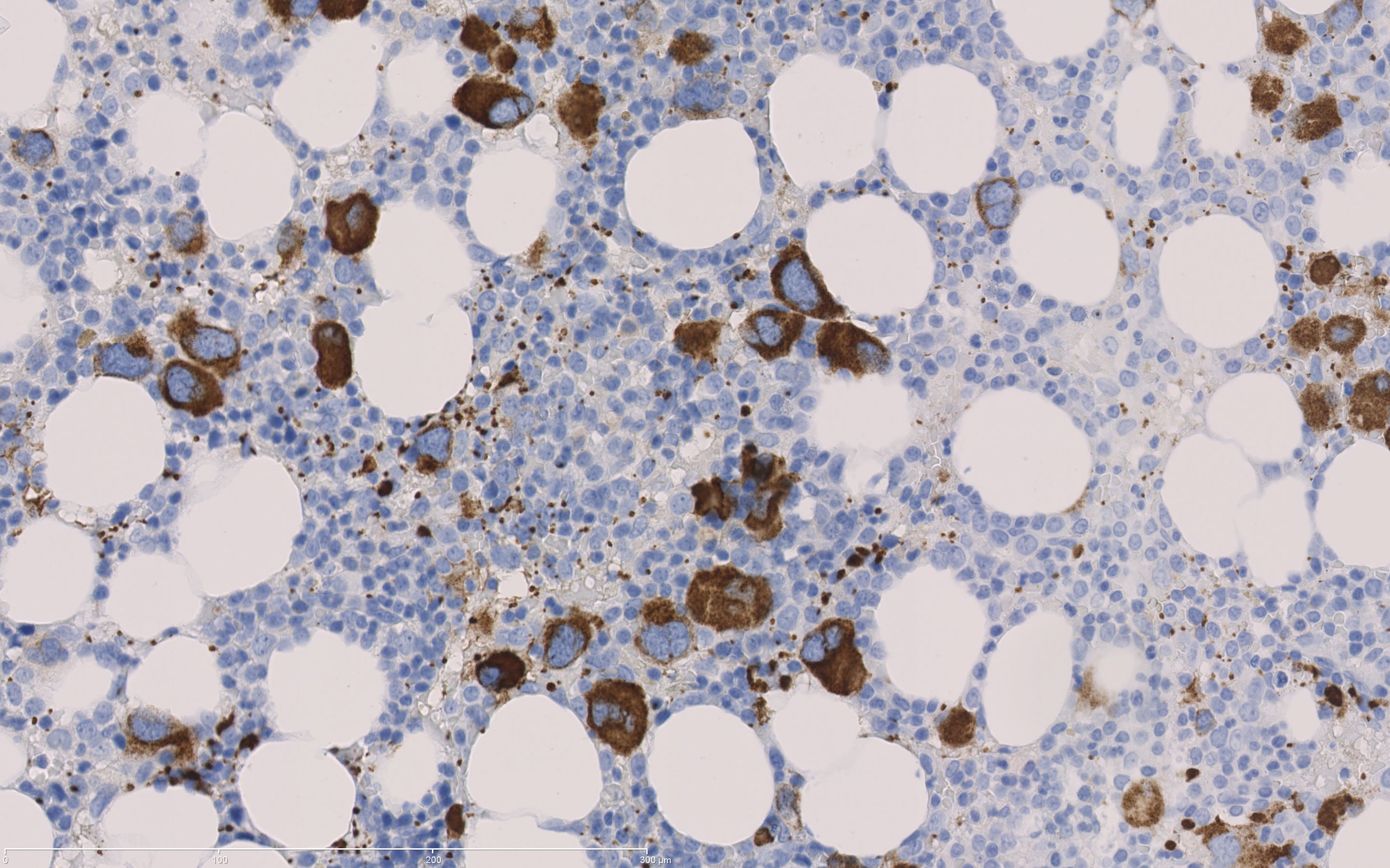
CD061 positive megakaryocytes
|
| CD068 | (cytoplasmatisch) Tonsil: Granulocyten en macrofagen in de kiemcentra zijn matig tot sterk gekleurd | 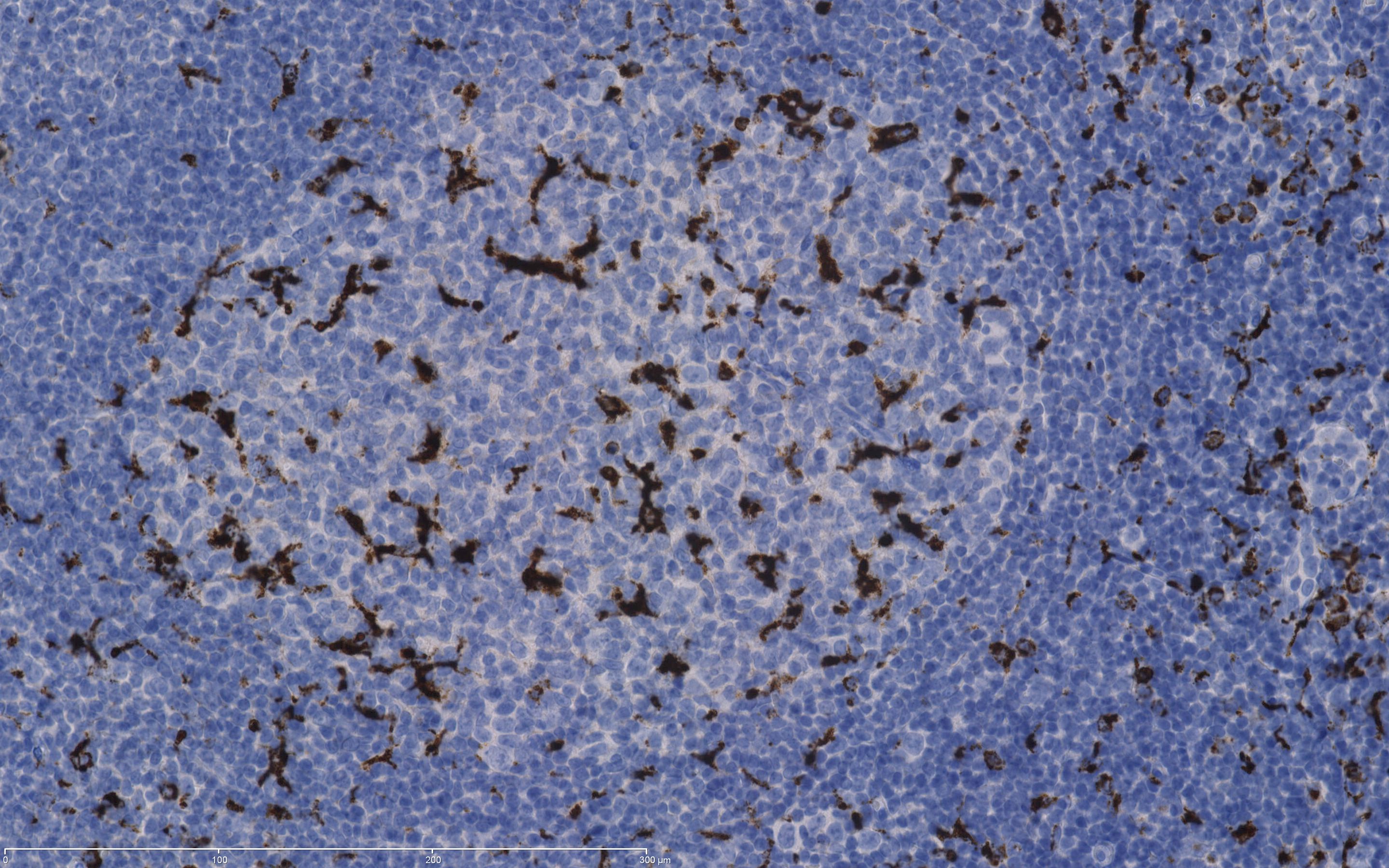
Tonsil: CD 68 in granulocytes and germinal centre macrophages
|
| CD079a | (cytoplasmatisch en membraneus)Tonsil: Alle B-cellen inclusief die van de kiemcentra en mantel zone zijn matig tot sterk positief en de plasmacellen zijn sterk positief. | 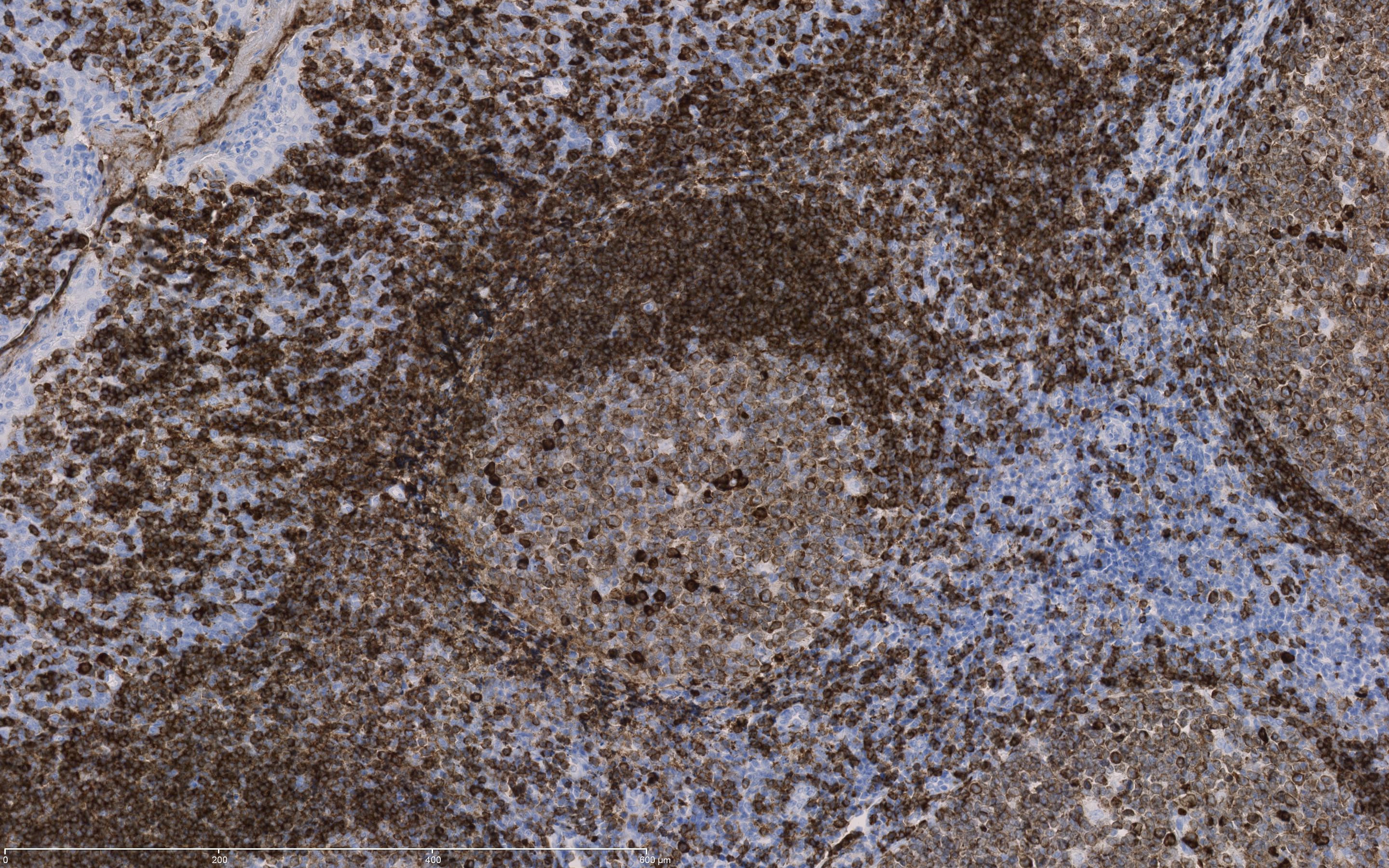
CD79a in tonsil: The mantle zone B-cells show an intense membranous staining, while the germinal centre B-cells show a moderate staining. Plasma cells and late stage germinal centre B-cells show a strong cytoplasmic staining
|
| CD099 | (membraneus en cytoplasmatisch): de basale en para basale laag van het plaveisel epitheel kleurt vooral membraneus matig tot sterk aan | 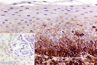
Tonsil: the basal and parabasal squamous epithelial
|
| CD117 | (cytoplasmatisch, membraneus) Appendix: mestcellen en cellen van Cajal zijn matig tot sterk aangekleurd, spier moet negatief zijn | 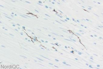
Appendix CD117 positive Cajal cells
|
| CD138 | Tonsil: Plasma cellen en plaveisel epitheel zijn CD138 positief. | 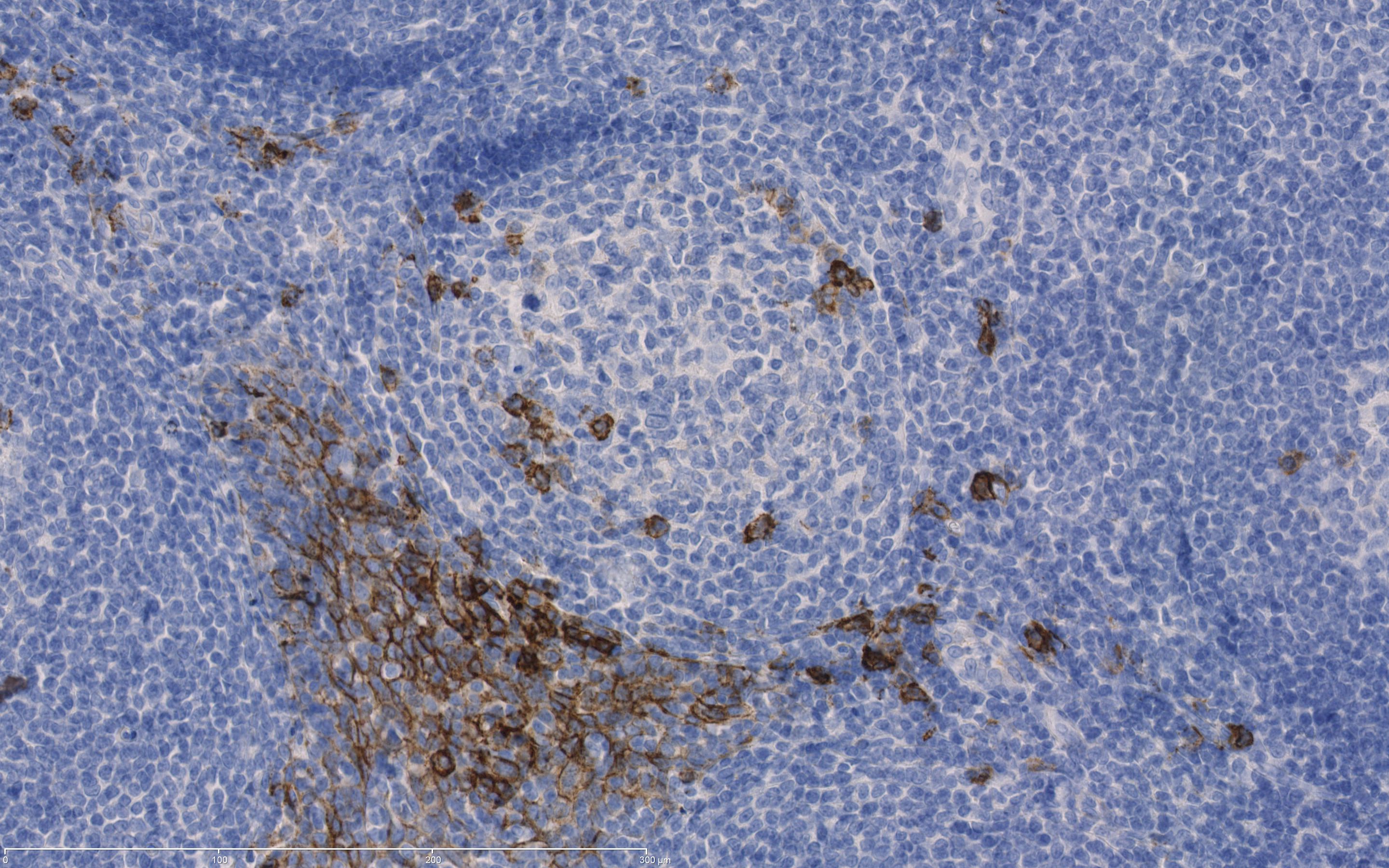
Tonsil: Plasma cells and squameus epithelium are stained.
|
| CD19 | (membraan, cytoplasmatisch) Tonsil: praktisch alle B cellen dienen te zijn aangekleurd. Tcellen dienen negatief te zijn. | 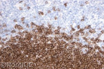
CD19: normal tonsil: a strong and distinct membranous staining reaction is seen in virtually all B-cells. T-cells are negative
|
| CDX2 | (kern) Appendix: alle kernen van het epitheel zijn matig tot sterk positief ( de basaal cellen moeten sterk aankleuren de rest van de cellen matig). Pancreas: is een betere controle dan appendix. Epitheel van de grote buizen dient positief te zijn. |

CDX2 in Appendix/pancreas: All nuclei of columnar epithelium are showing moderate (luminal cells) to strong (basal cells) staining reaction/pancreas: large ducts has to show a weak to moderate positivity
|
| CEA of CD66e (Carcinoembryonic Antigen) | (cytoplasmatisch) Appendix: CEA wordt matig tot sterk tot expressie gebracht door het oppervlak (grenzend aan het lumen) van de epitheel cellen, de rest van het cytoplasma is in mindere mate positief. Lever is negatief. | 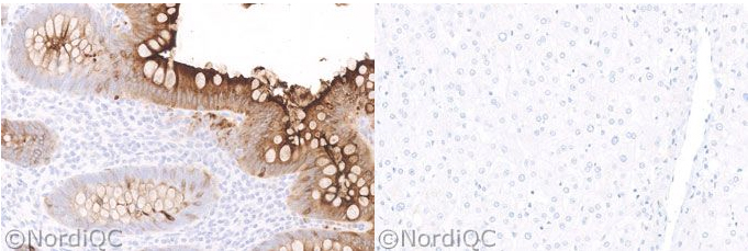
Appendix/Liver: epithelial cells show a moderate to strong cytoplasmic staining reaction. At the luminal surface the staining is stronger/ Liver is negative
|
| Chromogranin-A | Appendix: neuroendocriene cellen in het epitheel tonen een sterke aankleuring en de perifere zenuwen, ganglion cellen en axonen (inclusief plexus van Auerbach) tonen een matige cytoplasmatische aankleuring. De spiercellen moeten negatief zijn. | 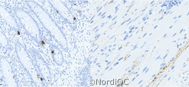
Appendix: both the axons of the peripheral nerves and the ganglion cells show a weak to moderate distinct granular staining reaction, while the smooth muscle cells are negative. Neuro-endocrine cells in the mucosa are strong positive.
|
| cker pan (MNF16, KL1, AE1/3) | Lever: Galgang epitheel is sterk cytoplasmatisch aangekleurd. De lever cellen laten matige vooral membraneuze aankleuring zien. |
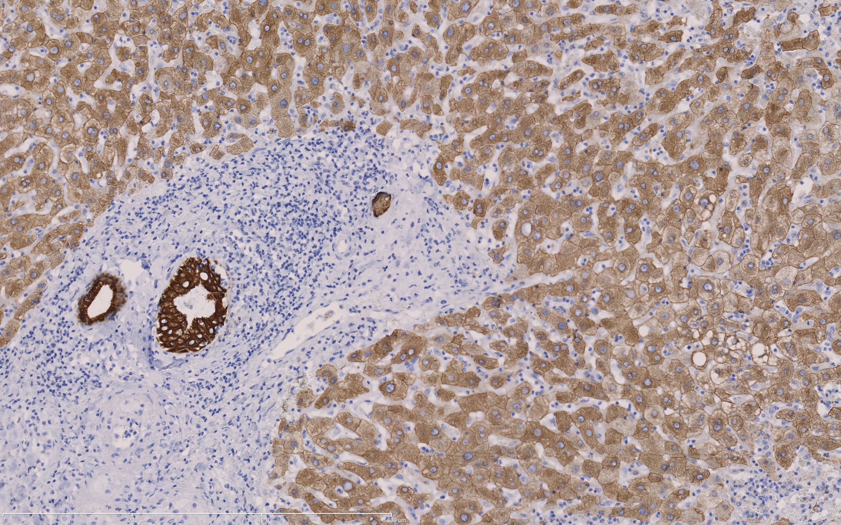
Liver: hepatocytes are moderate positive, gall epithelial is very strong positive
|
| CMV | (kern en cytoplasmatisch) De MCMV-geinfecteerde cellen zijn positief. | 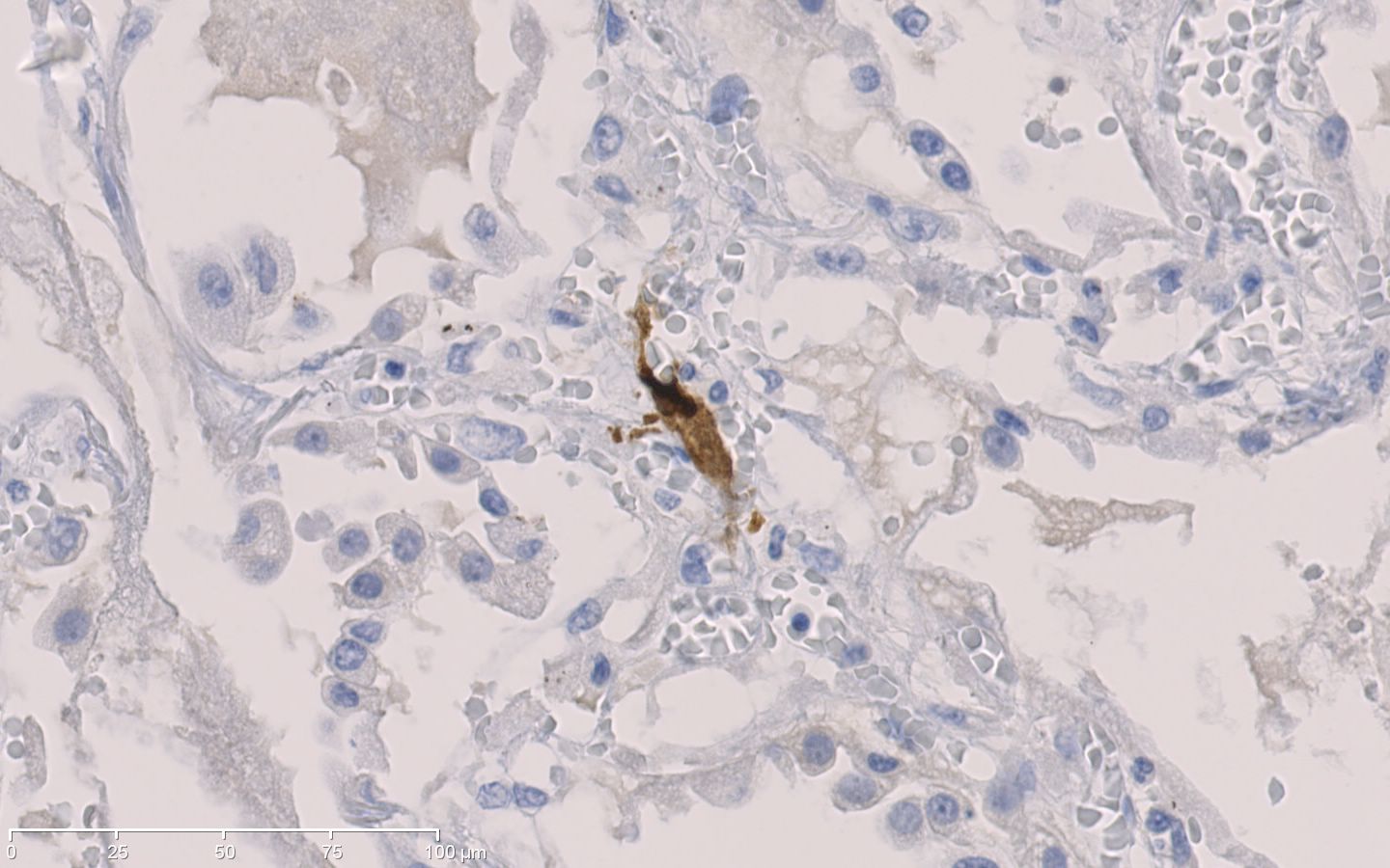
De MCMV-infected cells.
|
| Collagen III | (cytoplasmatisch) Lever: sinusoiden en het extracellulaire matrix zijn matig tot sterk aangekleurd, hepatocyten en de portale velden zijn negatief. Basaal membranen van de bloed en lymfevaten, de galgangen zijn positief. | 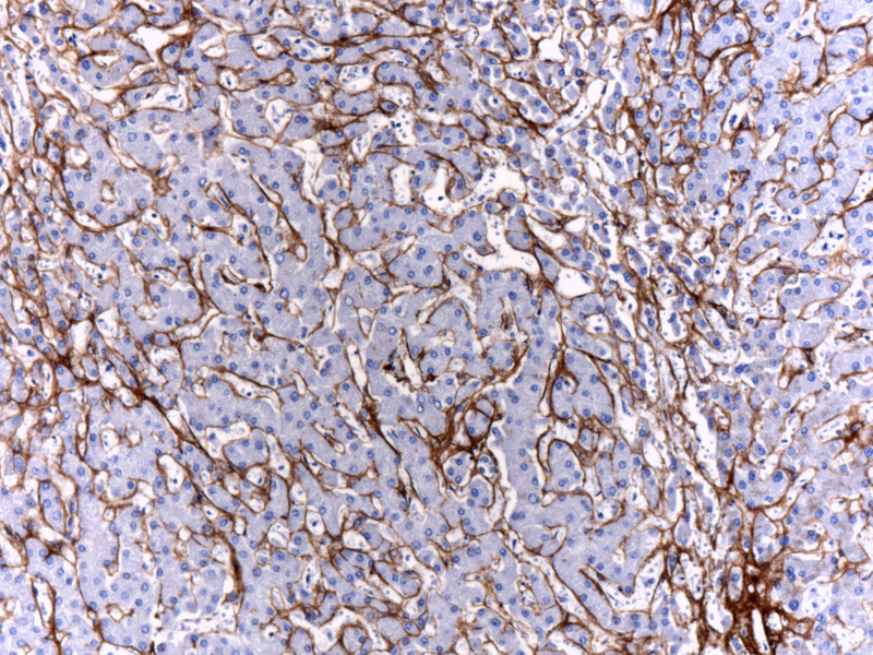
Liver: Collagen III in hepatic sinusoids
|
| Collagen IV | (cytoplasmatisch) Lever: sinusoïden zijn matig tot sterk aangekleurd, hepatocyten en de portale velden zijn negatief. Basaal membranen van de bloed en lymfevaten, de galgangen zijn positief. |
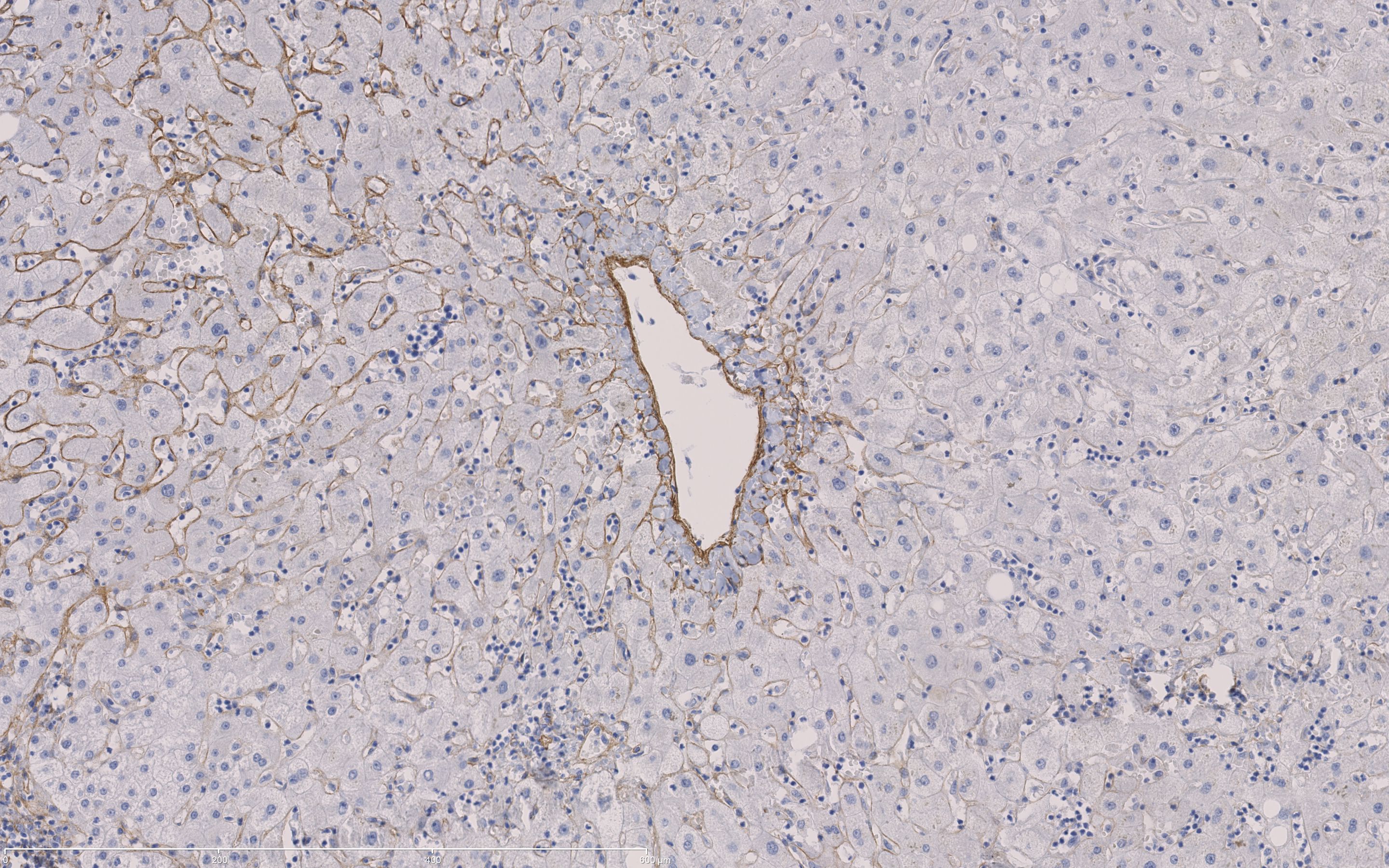
Liver: peri sinusoids are expressing COL IV
|
| Cyclin D1 | (kern) Tonsil: Parabasale laag van het plaveiselepitheel kleurt positief aan, zo ook endotheel en een paar fibroblasten. |
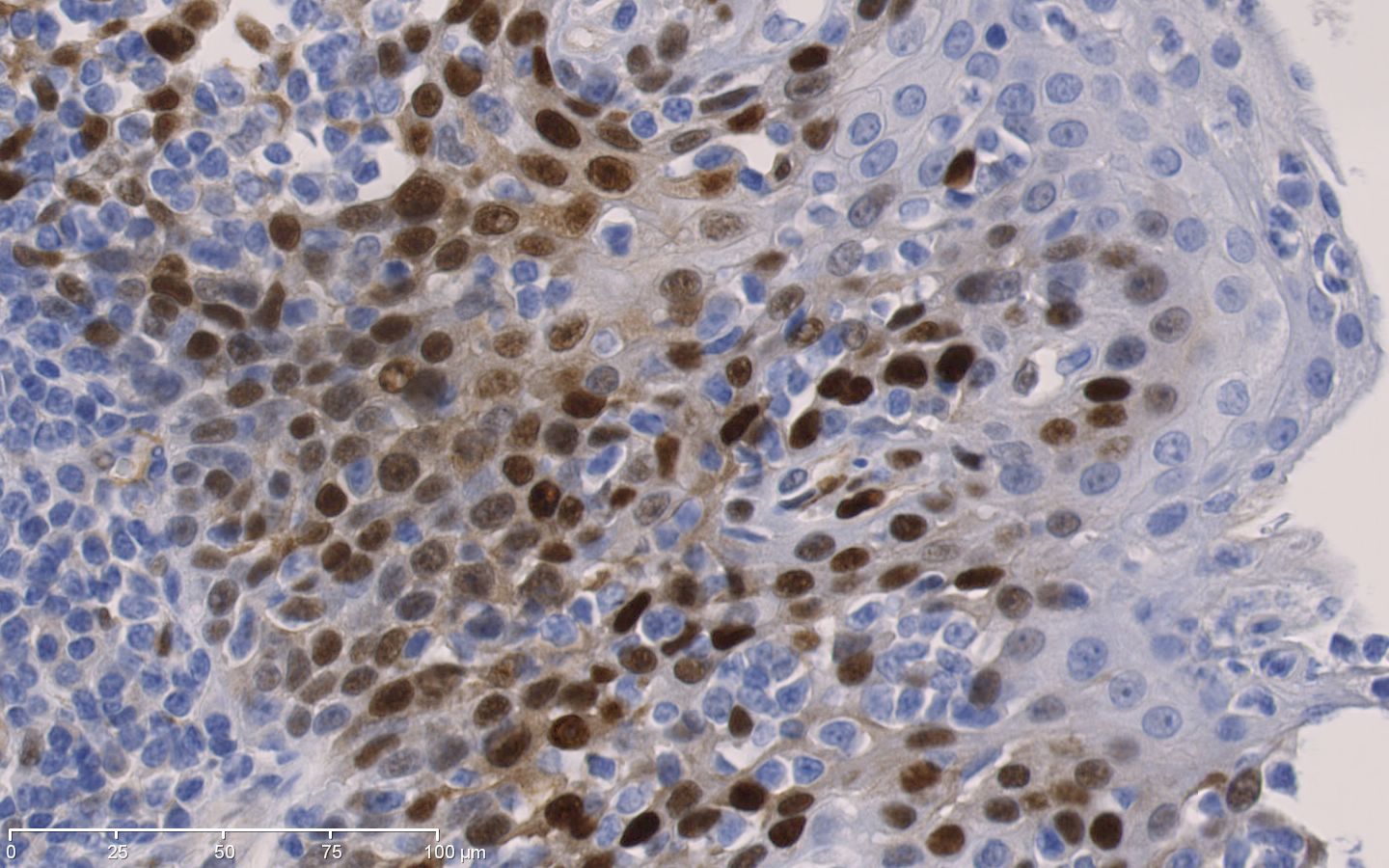
Tonsil (nuclear):Parabasal squamous epithelial cells are positive
|
| Cytokeratin 05 | Tonsil: Het plaveisel epitheel is positief in alle cel lagen. | 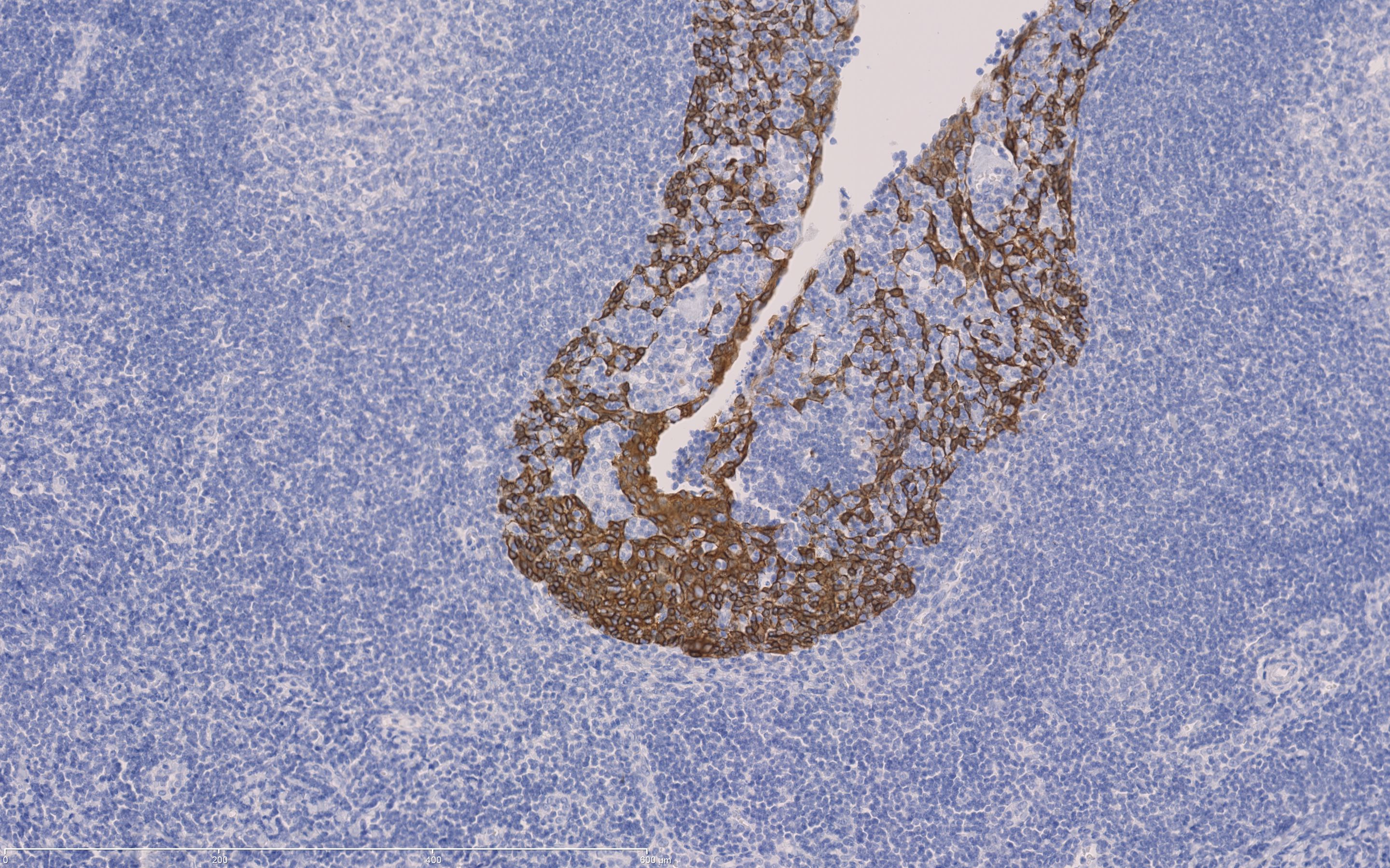
CK 5 in tonsil: Virtually all the squamous epithelial cells show a distinct, moderate to strong cytoplasmic staining, while no background staining is seen
|
| cytokeratin 07 | Pancreas: De epitheelcellen van de grote, middelmatige gangen zijn sterk positief terwijl de andere gangen matig tot zwak positief zijn. | 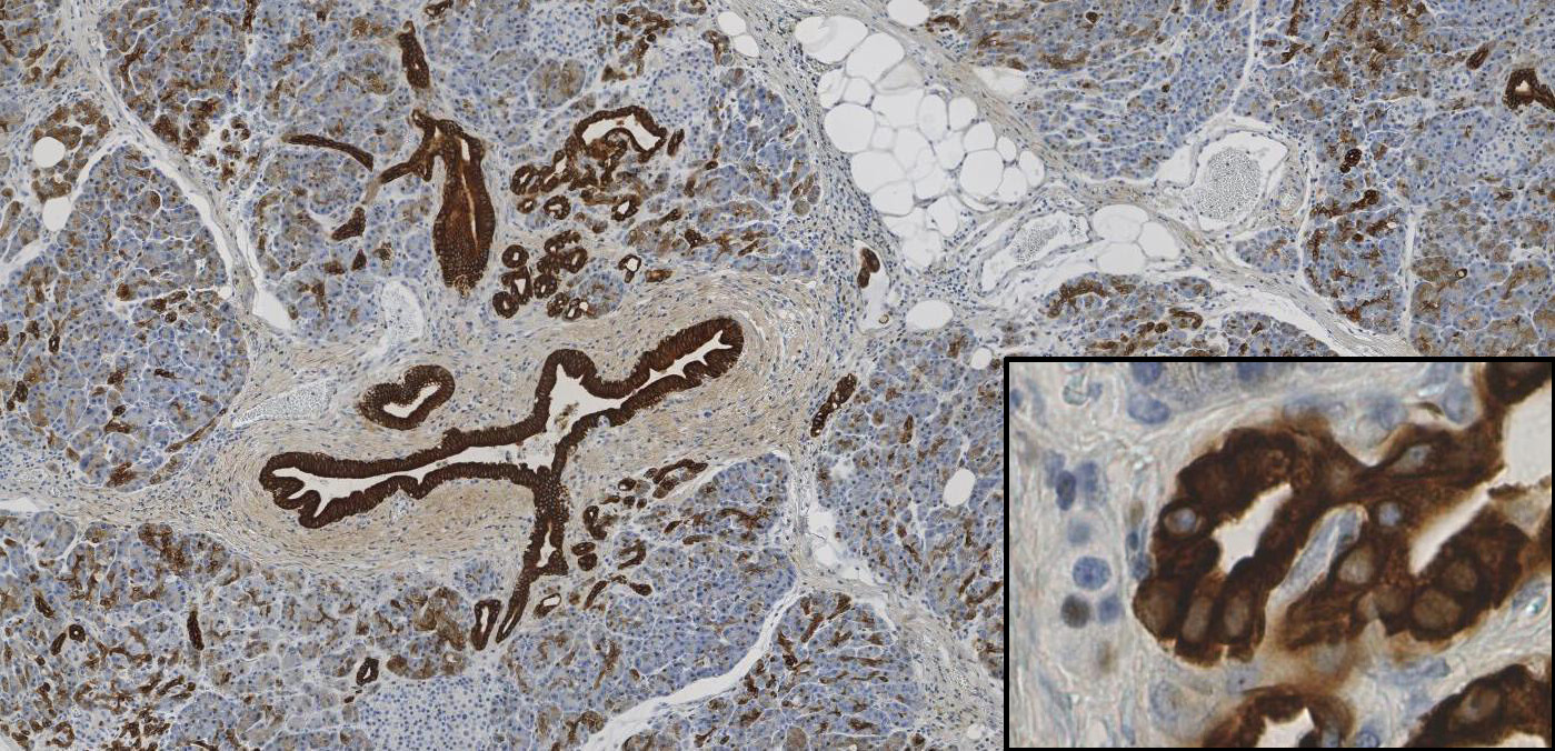
CK 7 in Pancreas: the epithelial cells of the large interlobular ducts show a strong cytoplasmic reaction, while the epithelial cells of the intercalated ducts a weak to moderate reaction
|
| Cytokeratin 10 | Huid: Suprabasaal plaveisel epitheel is positief, basale cellen zijn negatief | 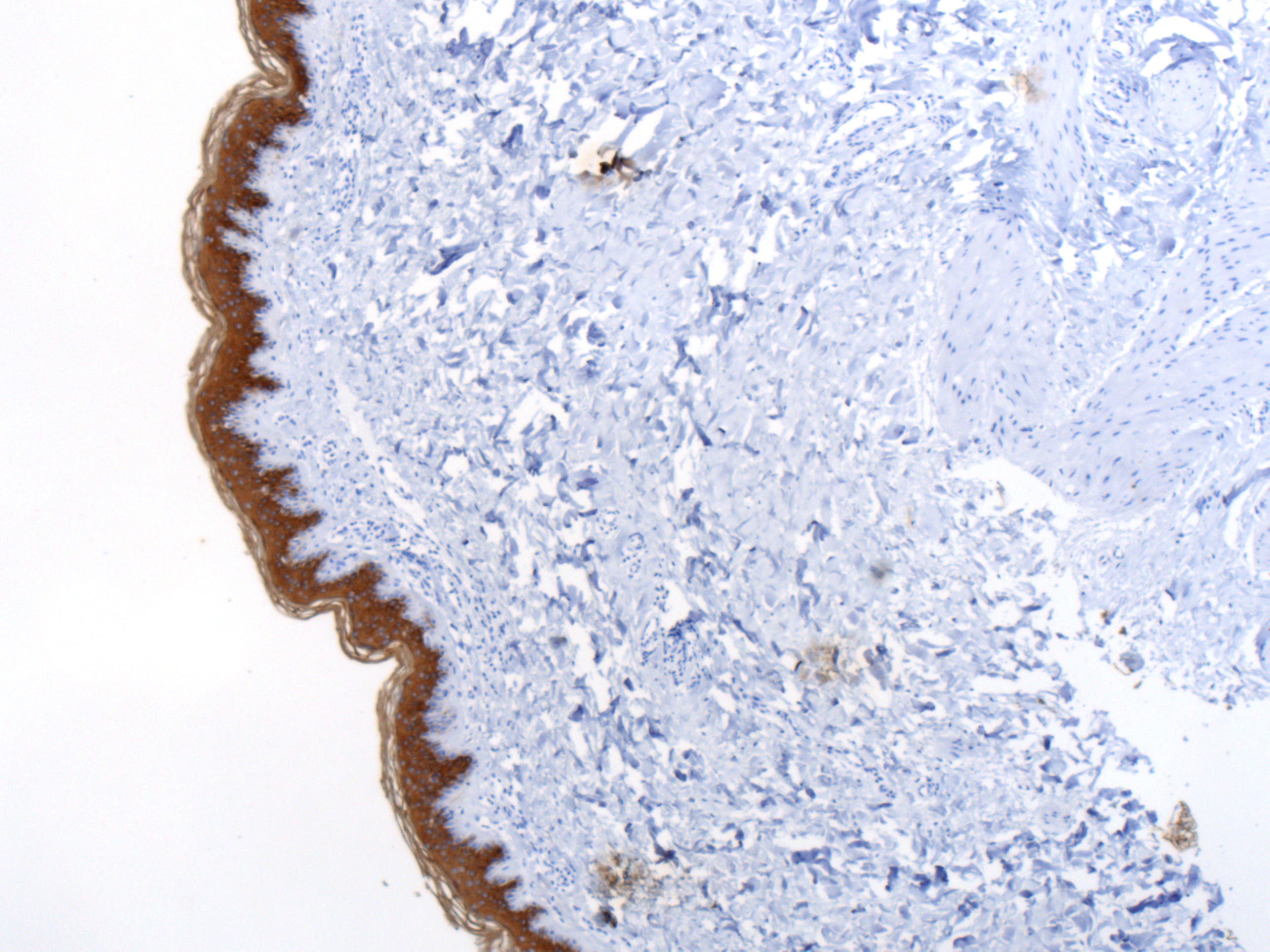
Skin:: Suprabasal squamousepithelial cells are positive.
|
| Cytokeratin 14 | Tonsil: plaveisel epitheel is ckeratine 14 positief | 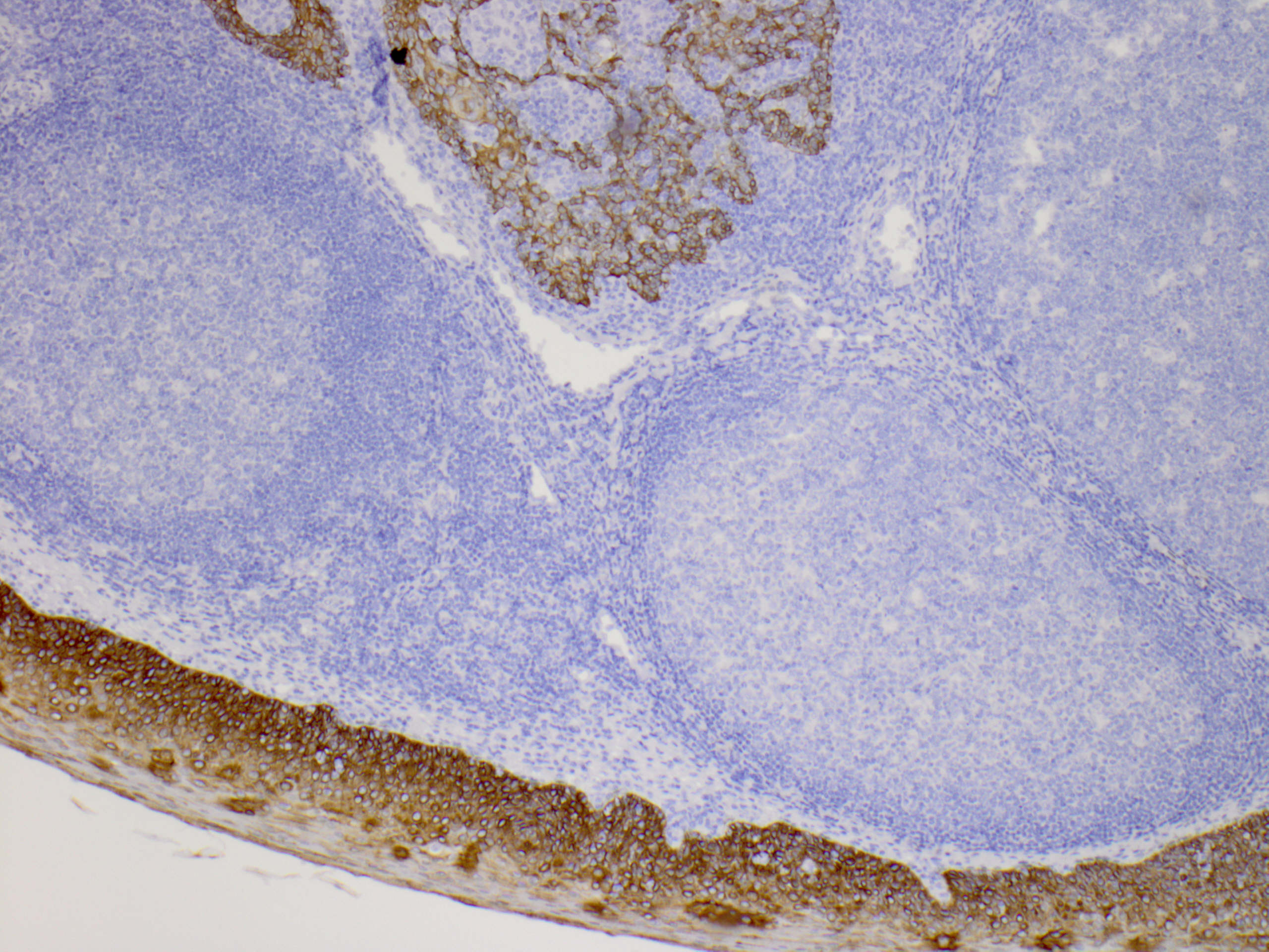
|
| Cytokeratin 18 | Lever: alle hepatocyten (over het algemeen een wisselende expressie) zijn zwak tot matig aangekleurd en galgang epitheel is heel sterk positief. |
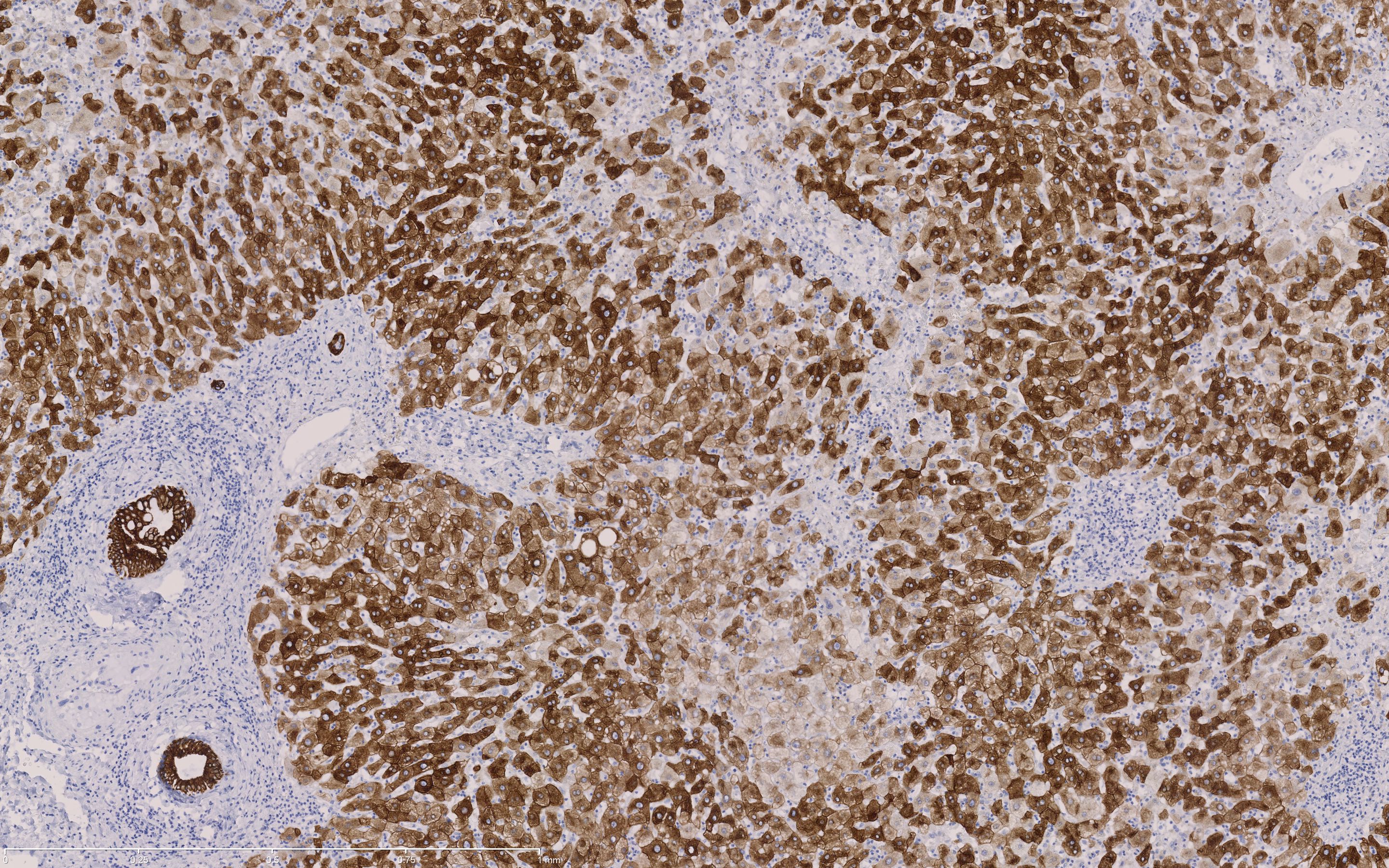
cker 8,18: The bile duct show a strong cytoplasmatic staining. Liver cells are weak to moderate stained.
|
| cytokeratin 19 | Appendix: epitheel en mesotheel is positief. Endotheel kan ook aankleuren. |
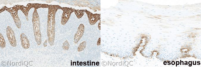
Appendix: The surface columnar epithelial cells show a strong cytoplasmic staining reaction, while the columnar epithelial cells in the basal parts of the crypts show a weak to moderate staining reaction
|
| Cytokeratin 20 | Appendix: De cellen die grenzen aan het lumen laten een sterke aankleuring zien terwijl de cellen in de basale crypten negatief tot zwak zijn aangekleurd. Neuroendocriene cellen zijn matig tot sterk aangekleurd. | 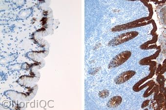
Left: Gastric mucosa: The majority of the foveolar epithelial cells show a distinct cytoplasmic reaction.
|
| cytokeratin HMW (34b12) | Prostaat: Ker HMW is positief in het cytoplasma van basale cellen in normale klierbuizen. |
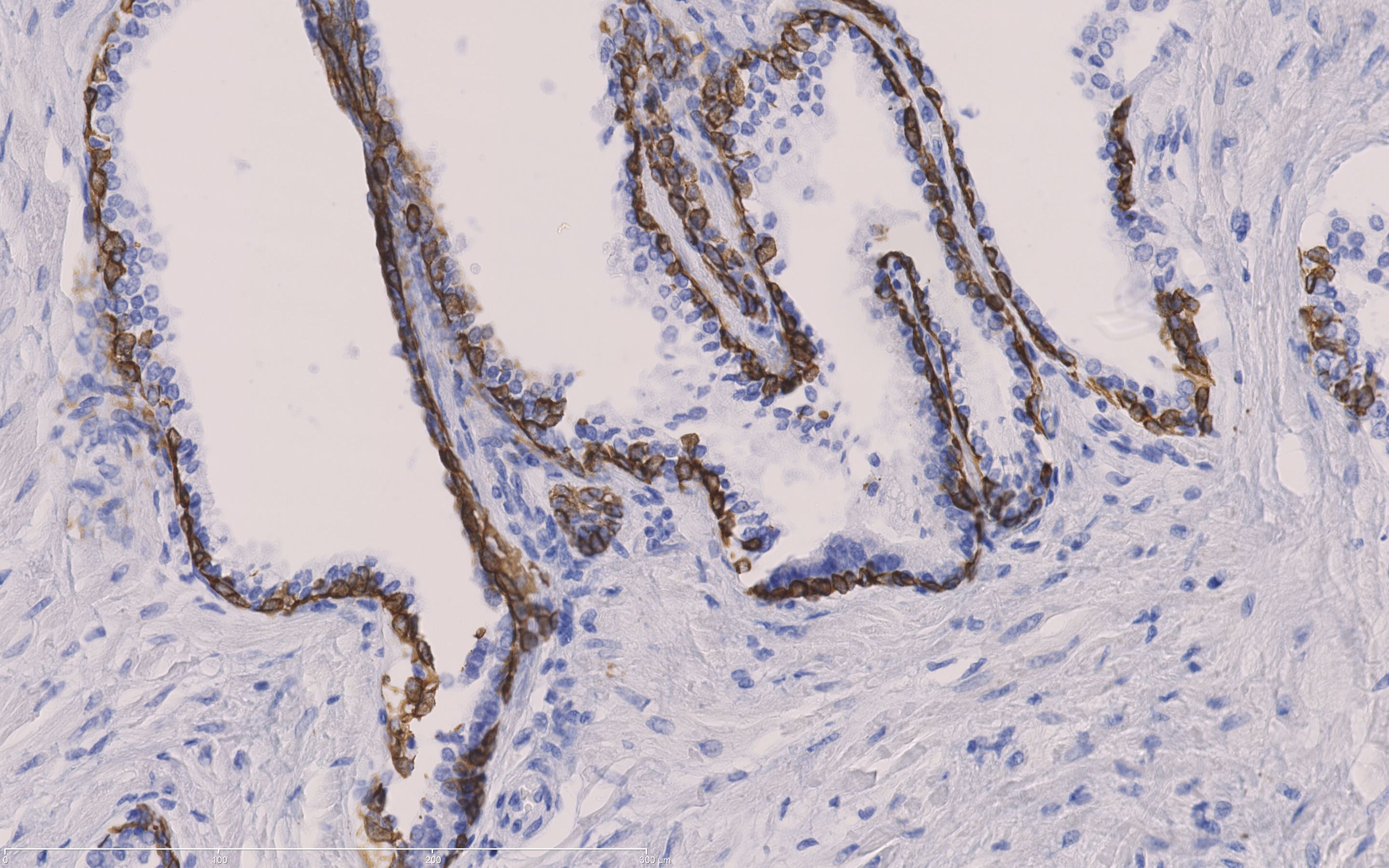
Prostate: Cytokeratine high moleculair weight
|
| cytokeratin pan (AE1/3) | Lever: Galgang epitheel is terk cytoplasmatisch aangekleurd. De lever cellen laten matige vooral membraneuze aankleuring zien. | 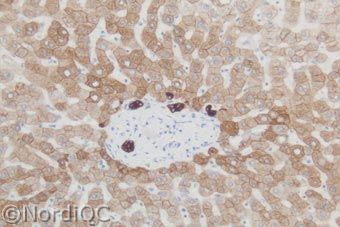
Liver: the majority of the liver cells are positive. The bile ducts show an even more cytoplasmatic reaction.
|
