Description
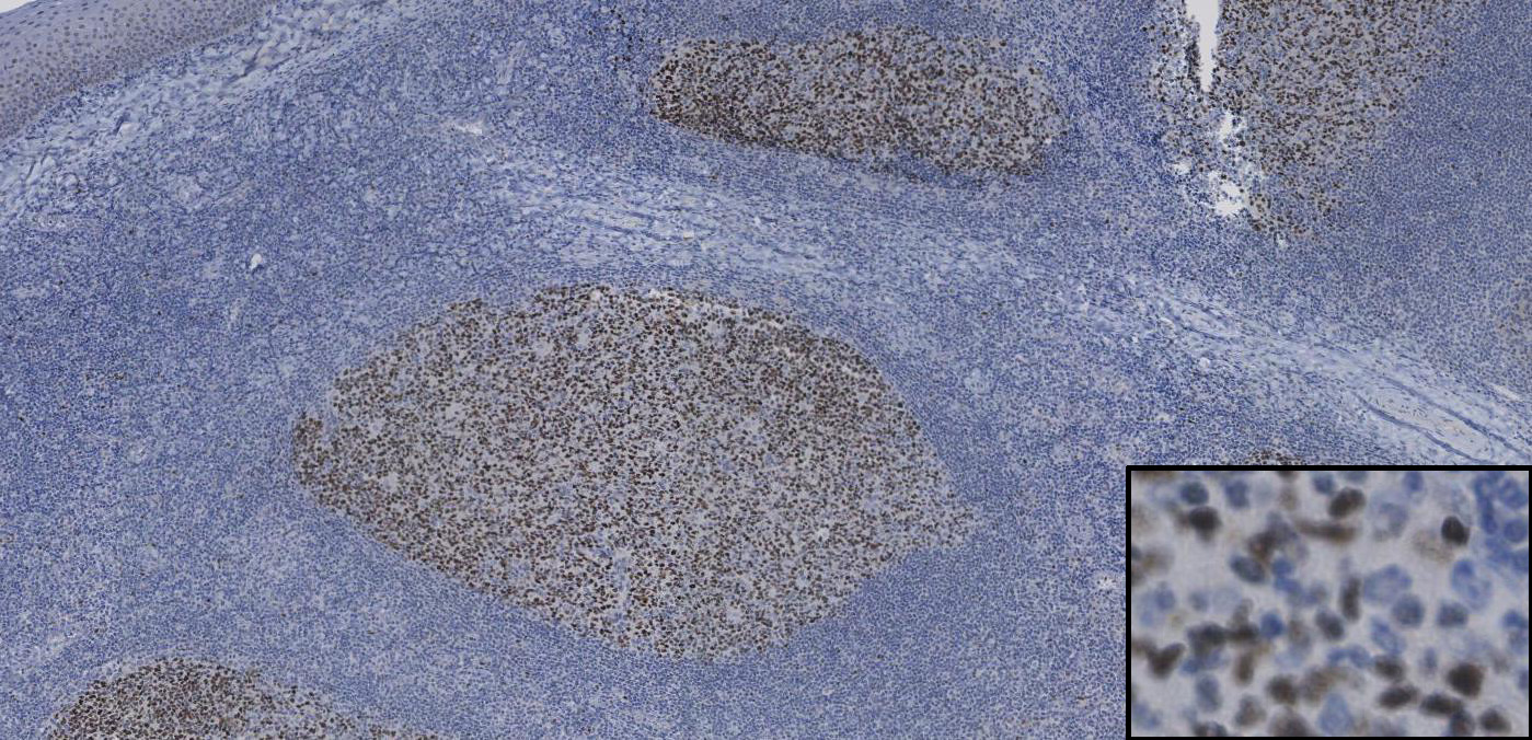
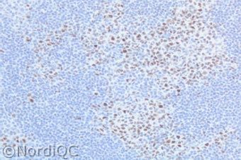
(Kern) Tonsil: sterke kernaankleuring van de B-cellen in het kiemcentrum en een zwak tot matige aankleuring van de kernen van het plaveisel epitheel.
Additional information
(kern) Appendix: in het kiemcentrum van de B-cellen, epitheel kan zwak positief zijn.
Wordt gebruikt voor de classificatie van diffuus grootcellige lymfomen , folliculaire lymfomen en het Burkitt lymfoom.
In maligne lymfomen, wordt BCL-6 het meest gedetecteerd in afwijkingen afkomstig van het kiemcentrum zoals bijvoorbeeld het folliculaire lymfoom, het Burkitt lymfoom, sommige diffuus grootcellig B-cel lymfomen, het nodulaire lymfocyten predominante Hodgkin lymfoom en zelfs in het T-cel lymfoom met een folliculaire lokalisatie (folliculair T cel lymfoom). Het wordt ook aangetroffen in het CD30+ anaplastische grootcellig lymfoom.
BCL6 expressie wordt niet aangetroffen in acute lymfatisch leukemie, in marginale zone lymfomen en het mantel cel lymfoom.
Voor meer informatie/referenties klik
Nordiqc en
hier en
hier en
hier en
hier en
hier en
hier en
hierVoor meer informatie over lymfomen klik
hier