BCL-2

  • image

    Tonsil: The scattered T cells in the germinal centers and the mantle zone Bcells are moderate to strong positive

  • image

  • image

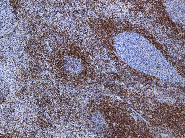
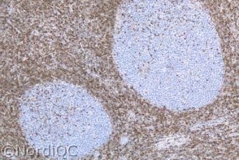
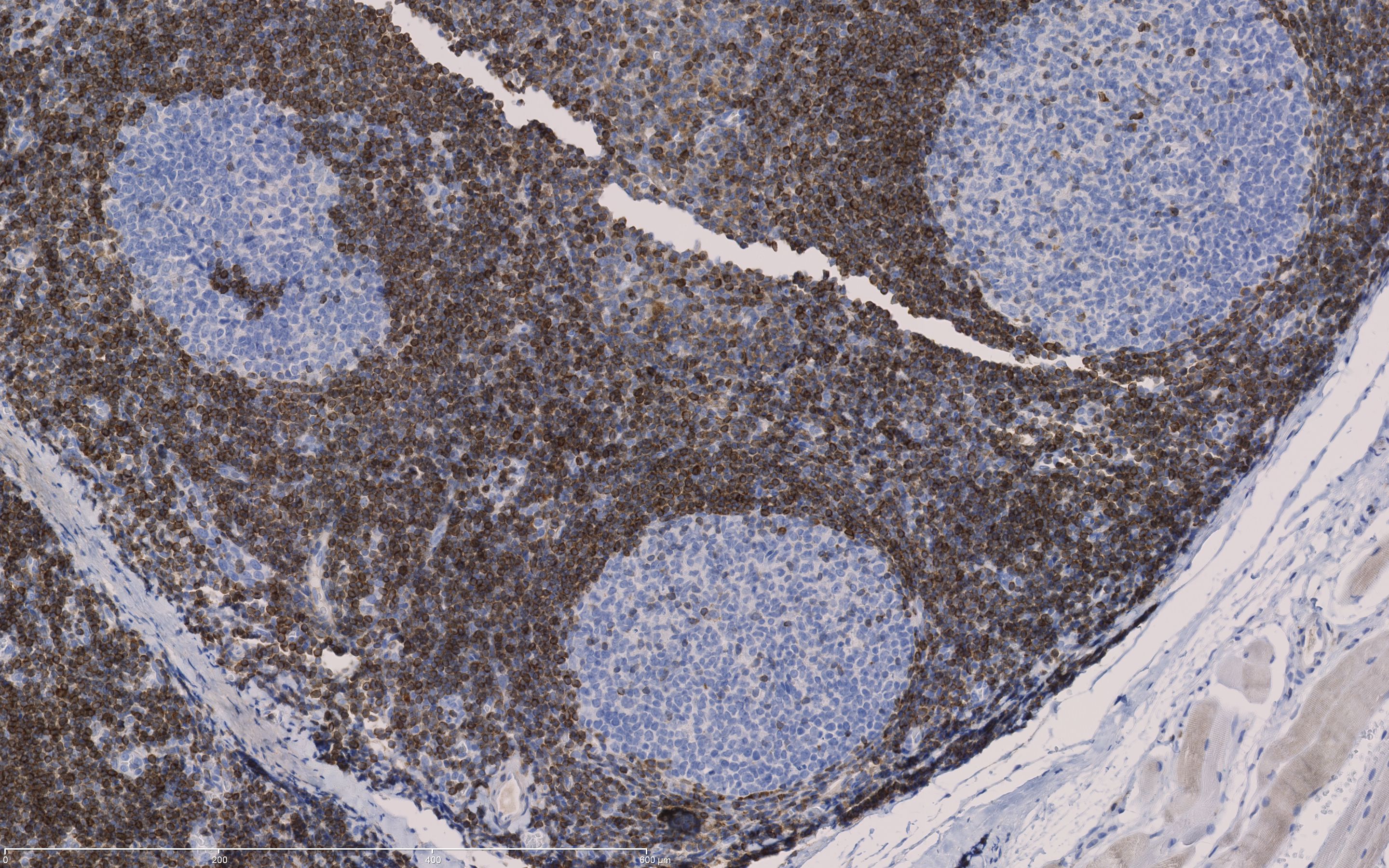
    BCL2: Tonsil: Virtually all the peripheral B- and T-cells show a strong staining. In the germinal centres, scattered T-cells show a distinct staining, whereas the B-cells are negative

  • image

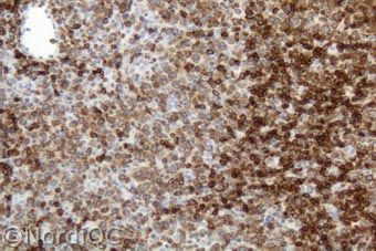
    BCL2: Bcl-2 staining of the follicular lymphoma grade III. Virtually all the neoplastic show a moderate staining, while the remnants of the normal lymphocytes (right) show a strong staining.

  • image
  • image
  • image
  • image
Description
Tonsil (cytoplasmatisch): alle perifere lymfocyten zijn positief, inclusief T-cellen in de follikels. B-cellen in de follikels zijn negatief. De basale laag van het epitheel wordt zwak tot matig aangekleurd.
Additional information
Appendix: Alle perifere lymphocyten incl T-cellen in de follikels zijn positief. B cellen in de kiemcentra zijn negatief. Pancreas: zwakke aankleuring van het epitheel.

Wordt primair toegepast bij de diagnostiek van het folliculair lymfoom (BCL2 positief) versus een reactieve secundaire (BCL-2 negatieve) follikel en het uitsluiten van een Burkitt lymfoom (BCL2 meestal negatief of soms een zeer zwakke aankleuring) bij de differentiaal diagnose Burkitt lymfoom vs diffuus grootcellig B-cel lymfoom (BCL2 positief).

Overexpressie van BCL2 komt voor in vele typen kanker, inclusief NHL, leukemie, adenocarcinoma, plaveiselcelcarcinoom, verschillende type sarcomen, neuroblastomen etc. Sterke expressie van BCL2 wordt gevonden in gastro intestinale tumoren, solitaire fibreuze tumoren en het synovial sarcoom. Terwijl fibromatosis en het maligne fibro histiocytoma meestal negatief zijn.

Voor meer informatie/referenties klik Nordiqc en hier en hier en hier en hier en hier en hier en hier en hier

Voor achtergrond van lymfomen klik hier