Description
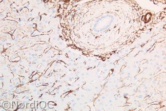
Appendix: gladspierweefsel in vaten en spierlagen. Myofibroblasten onder de epitheel laag kleuren aan.

Liver: alpha-SMA he smooth muscle cells in the portal vessels as well as the perisinusoidal smooth muscle cells show a distinct staining. The liver cells are negative
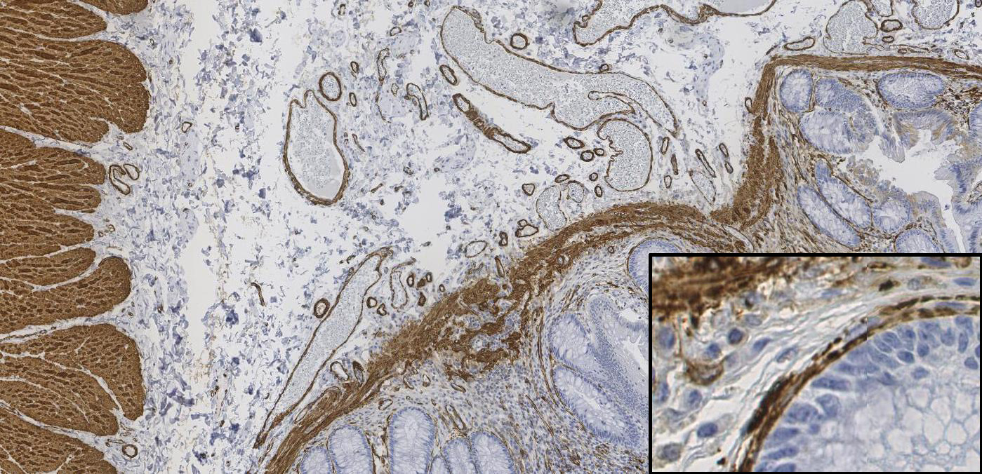
Appendix: clone 1A4) of the normal appendix. Intense staining is seen in the smooth muscle cells of the lamina muscularis mucosae. More important the tiny layer of myofibroblasts lining the surface epithelial cells is stained.